Total Hip Replacement (THR)/ Hip Arthroplasty
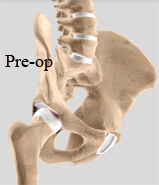
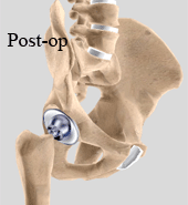
Hip Joint replacement or Total Hip Replacement is surgery to replace all or part of the hip joint with an artificial device to restore joint movement (prosthesis).
There are different types of hip replacements. If a hemi-arthroplasty is performed, either the femoral head or the hip socket (acetabulum) will be replaced with a prosthetic device. In a total hip replacement, both the femoral head and the hip socket is replaced by the prosthetic device.
WHAT IS ARTHRITIS AND WHY DO JOINTS WEAR OUT?
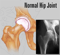
The normal joint in our body is made up of two bones which are lined by surface cartilage. The joint is surrounded by a capsule which has a thin lining of synovial cells which produce a thin layer of lubrication film. The lubrication film (synovial fluid) together with the surface cartilage (articular cartilage) acts as a shock absorber and allows the joint to move smoothly and lasts for many, many years.
If the surface cartilage is badly damaged or if the joint surfaces are not aligned properly (example, in a shallow hip) then the cartilage will wear out much more quickly than the normal wear and tear and as a result the bone under the cartilage layer is exposed. The exposed bone starts to rub against each other and the process of osteoarthritis (wear and tear) is established.
Osteoarthritis is therefore the result of mechanical wear and tear on a joint. The main feature is a loss of surface cartilage with bone rubbing on bone. This process produces pain. The body tries to relieve this pain by increasing the amount of fluid in the joint. This is why joints are sometimes swollen. The formation of bone spurs and cysts around the joint is another hallmark of osteoarthritis.


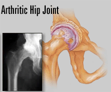
In an arthritic hip
The cartilage lining is thinner than normal or completely absent. The degree of cartilage damage and inflammation varies with the type and stage of arthritis.
The capsule of the arthritic hip is swollen
The joint space is narrowed and irregular in outline; this can be seen in an X-ray image.
Bone spurs or excessive bone can also build up around the edges of the joint.
The combinations of these factors make the arthritic hip stiff and limit activities due to pain or fatigue.
Diagnosis
The diagnosis of osteoarthritis is made on history, physical examination & X-rays
There is no blood test to diagnose Osteoarthritis (wear & tear arthritis)
Surgical procedure


You are admitted to the hospital and after appropriate pre-operative tests and admission procedures you will be taken to the operating theatre. The anaesthetist will discuss with you the type of anaesthetic. Anaesthesia may be either general or regional. With a general anaesthetic you are asleep and with a regional (spinal or epidural) your legs and hips are numb allowing you to have the operation without pain. Usually the anaesthetist will either sedate you or give you a full anaesthetic if you have a spinal/epidural procedure.
Most approaches to the hip are done with the patients lying on their side. When you are asleep you are positioned in a special brace that stabilises your pelvis and keeps you on your side. An incision is made along the side of your hip joint and the muscles carefully split and divided to expose the hip joint.
The worn out joint is exposed and the femoral head is resected. This allows visualisation of the acetabulum (socket). The socket is then cleared of debris and a reamer is inserted to appropriately fashion the socket to accept the artificial acetabular component.
After reaming is complete, the artificial socket is inserted. There are two types of sockets, (a) a cemented socket or (b) an uncemented socket. A cemented socket is cemented into the bone and an uncemented socket allows bone to grow into it. Your surgeon will advise you which is the most appropriate socket for your bone quality.
An uncemented socket has the ability to accept a socket lining which is either polyethylene (special plastic), ceramic or metal. The liner is inserted into the socket. Ceramic and metal articulating joint surfaces have lower wear rates than plastic sockets and therefore tend to be used in younger patients. The newer plastics last a lot longer than the older ones and are appropriately used in older patients.
After preparation of the socket, the femoral bone is prepared with various instruments to accept either a cemented or an uncemented femoral component. Once the canal is prepared the femoral stem is inserted with or without cement. A trial femoral head is placed on the stem and the hip is reduced. During the trial reduction the hip is tensioned appropriately and put through a range of motion. At the same time leg lengths and stability are examined.
Following the trial reduction the appropriate head is then placed on the stem and the hip is reduced. Occasionally leg lengths may not be entirely equal in order to tension the hip appropriately and thereby prevent dislocation.
Following insertion of the components the wound is closed usually with absorbable sutures and a drain is inserted.
What about the bearing (articulating) surface?
When the first hip replacements were made 35 years ago, it was found that over time they started to wear out and loosen. The reason they wore out was that fine plastic (polyethylene) particles were released from the socket which caused a small inflammatory response. This inflammatory response around the prosthesis caused the bone to weaken and the prosthesis to loosen and therefore a revision was needed. Occasionally these inflammatory areas can become large cysts and structurally weaken the bone so that when the hips are revised extra bone is required to fill up these defects. This extra bone may be taken from the patient or may be allograft bone, which is bone that has come from a bone bank.
In order to reduce the amount of wear particles, newer technologies have evolved. This includes new polyethylene, ceramic on ceramic and metal on metal articulations. The wear rates of ceramic on ceramic and metal on metal are 10 to 100 times less than the original plastic material.
Newer technology surfaces are tested in a laboratory on hip and knee simulators. The tests are extremely encouraging but only time will tell if they prove to be as successful as laboratory tests show. Younger patients tend to have ceramic or metal articulations in the expectation that less wear will occur and the joint will last longer.
Post-operative care:
When comfortable the physiotherapist will get you up and start your rehabilitation. You will be shown exercises to strengthen the muscles of the hip joint and you will also be shown the positions that you may keep your leg in and positions that will avoid hip dislocation. Initially you may start with a walking frame but then you will progress to crutches and a walking stick. Depending on your surgeon’s preferences you will either fully or partially weightbear. The wound will have a waterproof dressing over it, which will allow you to shower. It is important to mobilise as soon as you are comfortable as this will prevent complications such as deep vein thrombosis and chest infections.
To help protect your hip for the first 6 weeks after your total hip replacement.
- Do not bend your operated hip more than 90. Don’t lean forward when sitting, to reach anything!
- Avoid sitting on low chairs, stools or toilets or in car seats where your knees are higher than your hips.
- You should avoid crossing your legs or putting your operated leg across the midline of your body
- You should avoid lying on the operated side but you may be able to lie on the opposite side with a pillow between your legs.
- Avoid driving
- Avoid Crossing your legs
- Avoid lifting heavy items
- Avoid heavy housework
- Avoid lying on the operated side
- Avoid reaching towards your feet to dry them, put on footwear etc
Risks of hip replacement surgery:
Any operation that requires a general anaesthetic has certain risks attached to the general anaesthetic. In addition, there are also small risks attached to spinal or epidural anaesthesia. These risks will be discussed in more detail with your anaesthetist but the chances of having a major anaesthetic complication in New Zealand are one in 40,000.
Anaesthesia complications
As anybody undergoes general or regional anaesthesia (epidural anaesthesia) there are always risks associated with it. The risks of course are magnified if you have abnormal general medical conditions in addition to your older age, which may have affected the functions of your vital organs such as heart, lungs and kidneys. Therefore a complete evaluation of those systems has to be performed before you are taken to the Operating theatre
Specific risks for total hip replacement include the following:
Deep vein thrombosis and pulmonary embolus: You are given medication (injections) to thin your blood and prevent these complications. Other measures include TED stockings and calf compressors.
Infection: Superficial wound infections may occur early on and deeper infections can occur at a later stage. The incident of infection is less than 1%. Infections are usually treatable with antibiotic treatment. You are given antibiotics before the operation and for the first two days to prevent infections from happening. Very rarely, if a joint has a deep infection that cannot be controlled with antibiotic therapy, the joint requires removal and a second joint re-implanted at a later stage.
Leg length discrepancy: It is not unusual for there to be up to 1cm leg length discrepancy following a Hip replacement. This is quite easily tolerated. The reason there may be a discrepancy is to ensure that the hip joint is appropriately tensioned so that it does not dislocate. Initially you may think that you have a longer leg but this is often due to muscle contracture which over time will loosen up and your leg lengths will even out.
Hip dislocation: The risk of hip dislocation is usually less than 1 or 2%. Provided the components are placed correctly and the appropriate post-operative precaution measures adhered to, it is unlikely that the hip will dislocate.
Fractured femur: Very rarely the femoral bone may fracture at the time of surgery and this is usually treated immediately. It is also uncommon to fracture following a total hip replacement unless you have been involved in a bad accident.
Loosening of the prosthesis: As mentioned, over time the prosthesis may loosen if the bone does not grow into it sufficiently or if the bearing surface wears out to produce areas around the prosthesis, leading to loosening. Should a prosthesis loosen, then it can be revised. If only the bearing surface wears out, then usually only the bearing surface requires revision which is a much smaller operation. Patients who have metal on metal articulating surfaces have a slightly higher metal iron level in their blood. This has been extensively researched over the past 30 years and there have been no increased incidents of cancer or any other problems.
Damage to nerves and vessels: It is unusual to damage any major nerves or blood vessels following a hip replacement. However nerve palsy can develop if the nerve is stretched during surgery. Those with hip dislocations from childhood are at higher risk of nerve injury.
Haematoma: Occasionally a bleed may occur around the hip joint following the operation that may require drainage.
Scarring: Some patients tend to scar more than others and it may be that the scar that you have will be quite thickened (keloid).
Long-term swelling: Occasionally the operated leg may remain a little swollen for a number of months but in general this tends to resolve.
Trochanteric bursitis: Occasionally following hip replacement surgery one can experience inflammation at the side of the hip joint which usually settles with either a cortisone injection or anti-inflammatories.
Joint stiffness: Very rarely extra bone can form around your hip joint which will cause it to stiffen up again (heterotopic ossification). This is usually painless but may cause some stiffness.
General advice after hip replacement surgery:
- You should have a regular check every two years with an x-ray.
- If you have had any major bowel, bladder or dental surgery, antibiotic cover should be given prior to the surgery.
- Metal prostheses can activate security alarms at airports.
